Rear Brain: Structure And Functions
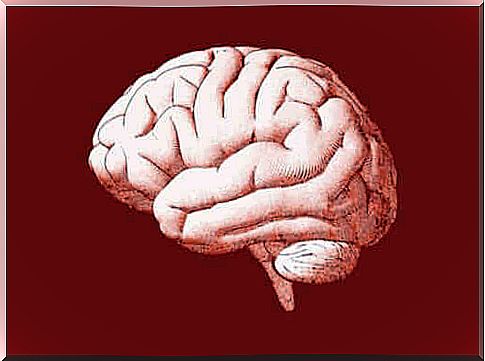
Throughout history, scientists have categorized the brain into several parts to try to better understand its functions and evolution. One of these parts is the posterior brain or rhombencephalon, which is an area that comes from the posterior primary embryonic vesicle.
The posterior brain is the posterior region of the brain. It is a structure formed by various sub-structures, which are responsible for several essential functions.
In this article we will show you the structure, differentiation processes and functions of this part of the brain.
The differentiation processes of the hindbrain
First, you need to understand the origin of the hindbrain . To be able to do this, it is important to explain what a differentiation process is. According to Bear, Connors, and Paradiso, authors of the book, Neuroscience: Exploring the Brain, it is a process through which structures become more complex and functionally specialized.
The first step in the differentiation process of the brain is the development of three primary brain vesicles of the neural tube, which originate from the anterior part.
The anterior part of the primary vesicles is the prosencephalon or better known as the forebrain. Then there is the vesicle located behind the forebrain called the mesencephalon or midbrain. And finally there is the back part of the vesicles, which are the hindbrain or rhombencephalon which are associated with the neural tube rear part.
The cerebrum is thus formed during an embryonic development through transverse swellings called rhombomeres, which are basically spaces that allow cells to form groups, each of which develops in different ways. In addition, they perform various functions.
The hindbrain has three essential structures:
- The cerebellum. It connects the brain stem with the brain bridge and it is a movement-controlling center that is fundamental to the body. It originates from the front part.
- The brain bridge. It is part of the anterior hindbrain, in front of the cerebellum and the fourth ventricle.
- Medulla oblongata. It is located in a position behind the cerebral bridge and cerebellum. It comes from the back.
In the vesicle stage, the anterior part of the hindbrain is shaped like a transverse tube. At the end, the rhombic lip or the dorsal-walled tissue of the tube grows forward and to the sides until it meets with the opposite side. The curl that is formed grows to form the cerebellum. And the ventral walls of the tube expand to form the brain bridge.
In differentiation of the posterior half of the hindbrain, some additional changes occur in the medulla oblongata. At one level, walling expands, leaving only the “roof” covered with ependymal cells instead of neurons. On another level, there is a system of white matter in the medulla oblongata along the ventral surface of each side.
Finally, with respect to the area of cerebrospinal fluid, it becomes the fourth ventricle, which continues the cerebral aqueduct of the mesencephalon.
The functions of the hindbrain
The cerebrum performs several functions:
- It is a fundamental part of the brain for information to pass through, from the prosencephalon to the bone marrow and vice versa.
- Its neurons take part in the process of sensory information.
- The neurons of the hindbrain contribute to voluntary movement control. In addition, they help regulate the autonomic nervous system.
- The cerebellum, which means “cerebellum”, regulates movements as if it were a control center. It also receives massive messages of axons coming from the bone marrow and the brain bridge. On the other hand, the cerebellum is responsible for comparing in-depth information and calculating the sequences of muscle contractions, which is essential for performing a movement.
- The medulla oblongata is responsible for carrying somatic information from the bone marrow to the thalamus. It also controls the movements of the tongue and is associated with sensory functions such as touch and taste.
- Auditory nerve axons are responsible for carrying information from the ear to the cochlear nuclei of the bone marrow. Then the nucleus projects axons to different structures: the tectum of the mesencephalon.
The in-depth information coming from the bone marrow also brings information about the spatial position of the body. The messages of the cerebral bridge carry information from the cerebral cortex and finally these messages specify the goal of the movement.
Possible health problems associated with the hindbrain
When the brain does not develop as it should, it could affect the hindbrain and its functions, which are vital for survival.
Here are some of the health problems associated with the hindbrain:
- Lesions in the posterior brain can cause movement problems, such as uncoordinated or inaccurate movements similar to those seen in people with ataxia.
- Damage to it can also cause deafness, for example, if there is an injury to the cochlear nucleus.
- Problems related to touch and taste.
- Dandy-Walker and Arnold-Chari malformations resulting from abnormal development of the hindbrain.
- An injured hindbrain can cause vomiting, weakness, breathing problems and circulatory problems.
- Rhombencephalitis, or inflammatory disease of the hindbrain, which can manifest itself due to many different factors.
The hindbrain plays an important role in the human body. Through its motor, sensory and visceral functions, it helps to regulate the human body. The consequences of its malfunction can greatly affect an otherwise healthy patient.
