Hippocampus: Structure And Functions
Cognitive processes such as learning and memory are vital for humans. The hippocampus plays a fundamental role in these processes, which is one of the areas of the brain.
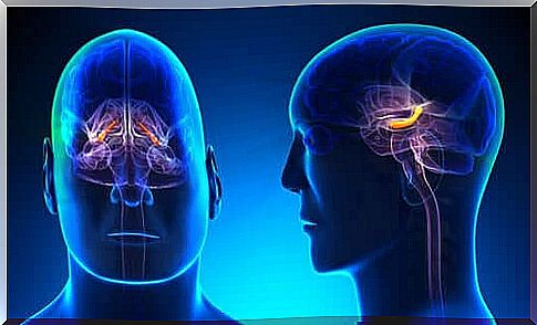
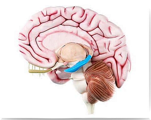
The hippocampus is a C-shaped prominent structure located in the lower part of the posterior ventricle of the brain. It consists of three primary parts (CA1-CA3).
Anatomical analysis of the hippocampus
In the 16th century, the anatomist, Arantius, mentioned the hippocampus for the first time. He named it the hippocampus, which comes from the Greek word for seahorse.
However, the area around the hippocampus also consists of the dentate gyrus, the subicular complex, and the entorhinal cortex.
All in all, the area is five centimeters long. In the middle part there is also the uncus, which is shaped like a potato and varies greatly from brain to brain.
The area around the hippocampus: Architecture
Dentate gyrus
The dentate gyrus is the middle part of the cerebral cortex. When it comes to cytoarchitectonics, the dentate gyrus is a trilaminate cortical area. The dentate gyrus has a typical C-shape and is ventrally separated from the first part of the hippocampus and from the subiculum at the fissure of the hippocampus.
The primary cell layer of the structure is full of granular cell bodies. The apical dendrites of these cells have branches in the dentate molecular layer. The granular cells and the molecular layers combined represent the fascia dentata.
The third innermost layer of the dentata gyrus is the polymorphic layer or hilus. Next to it, there is a fraction of the pyramidal cell layer covered with granular cells.
Hippocampus
The hippocampus has subregions called CA1, CA2, and CA3, which consist of a cell layer: the pyramidal cell layer. The surface that restricts the ventricular lumen, formed by axons of pyramidal cells, is called the alveus. Historically, this area consists of:
- Lucidum stratum
- Radiatum stratum
- Lacunosum molecular
Lucidum stratum, CA3, has fibers that form proximal dendritic synapses over the pyramidal cell wall in this layer. The CA2 layer is relatively compact and also presents a pyramidal cell layer, but its boundaries are difficult to define.
In addition, CA1 layer is a subarea in the hippocampus. The pyramidal cell layer in this region consists of an external and an internal layer.
Subiculum
The CA1 layer and subiculum overlap at the boundaries, forming a transition area. Subiculum is primarily divided into the following layers:
- Superficial: There is a broad molecular layer where subicular pyramidal cell dendrites are located. This pyramidal cell layer can be divided into two substrates: external and internal.
- External layer cells have a lipofuscin pigment accumulation in its apical dendrites.
- The presubiculum consists of superficial layers that contain modified pyramidal neurons.
- The parasubiculum has a cell layer that is difficult to differentiate from the presubiculum.
Entorhinal cortex
The term “entorhinal cortex” is a synonym for the Brodmann area. It extends primarily forward toward the middle portion of the tonsil nucleus, and backward toward the anterior commission of the transverse geniculate nucleus.
This area is in a way different from the rest of the brain.
Connections in the hippocampus
Hippocampal inner cycle
The connection for information in this part of the brain follows a unidirectional and glutamatergic (stimulating) pathway, which is part of a closed cycle. In this inner chain of connections, the dentate gyrus is very important as it receives the most information, which is then transmitted to the entorhinal cortex.
External connections
The external cycle consists of:
- Multiple cortical arteries.
- The amygdaloid complex.
- The middle septal core.
- Thalamus.
- The supramammary nucleus.
- The monoaminergic nucleus in the brainstem.
This is obviously the way the hippocampus receives sensory information from a variety of cortical areas.
Cortical connections
These projections serve primarily to introduce sensory information.
Subcortical connections
Fimbriae and fornix form the classical efferent system of the hippocampus. In addition, there are also connections between the hippocampus and the tonsil nucleus.
Eventually, the connections produced between the hippocampus and the hypothalamus are established through the subiculum.
As you can see, the hippocampus is a complex collection of areas that include the hypothalamus. Although most research projects have studied animals, it seems clear that the areas described here are very similar to those in humans.